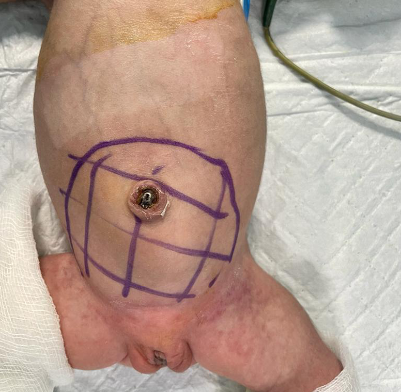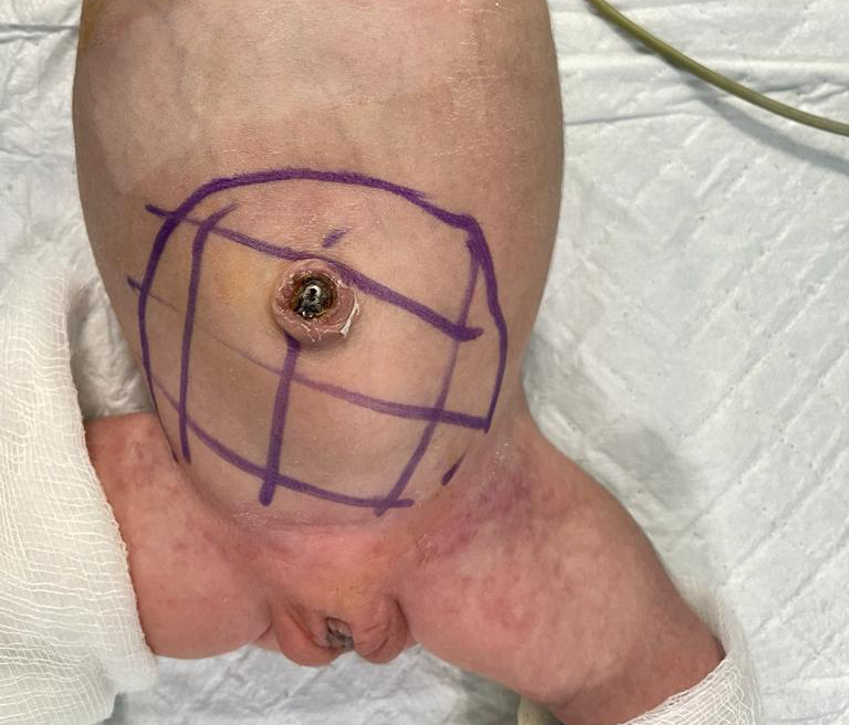
Sacrococcygeal Teratoma
The sacrococcygeal teratoma (SCT) is an unusual tumour that develops near the coccyx (base of the tailbone) in newborns. Babies with this birth abnormality are more likely to be females than males. Even though the tumours have the potential to become quite large, they are typically not malignant (i.e., cancerous). Although they occasionally cause issues before to delivery, they are typically curable by surgery.
SCT is typically identified by a sonogram because the uterus is larger than it should be or by a blood test on the mother at 16 weeks that reveals a high alpha fetoprotein (AFP) level. Polyhydramnios, a condition marked by an excess of amniotic fluid, frequently contributes to the uterus' enlargement. An ultrasonography examination can be used to determine SCT's diagnosis.